


Article reviewed by: Dr. Sturz Ciprian, Dr. Tîlvescu Cătălin and Dr. Alina Vasile
Article updated on: 22-05-2025
Lumbar spine bone edema – what it is, causes, symptoms and treatment
- What is lumbar spine bone edema?
- Causes and risk factors in lumbar bone edema
- Symptoms and manifestations of lumbar bone edema
- Diagnosis – how is lumbar bone edema detected?
- Treatment options for lumbar bone edema
- Hyperbaric oxygen therapy in lumbar bone edema
- Conclusions and advice for patients
Bone edema at the lumbar spine level is a condition that patients often hear about following an MRI examination, but the term can cause worry and confusion. In short, "bone edema" refers to fluid accumulation inside the bone, more precisely in the bone marrow of lumbar vertebrae. This change frequently appears as a sign of an underlying problem, rather than as a standalone disease. But, once we understand the causes and treatment method, bone edema can be managed efficiently, allowing amelioration of back pain and return to normal activities.
What is lumbar spine bone edema?
Lumbar spine bone edema represents abnormal fluid accumulation in bone marrow spaces, especially in vertebral bodies. This phenomenon indicates bone suffering, appearing following an inflammatory process, microfractures, or prolonged mechanical stress. On magnetic resonance imaging (MRI), edema appears as an area with increased signal on T2 and STIR sequences, suggesting fluid presence in deep bone structure.
Unlike classic soft tissue edema, bone edema is not visible externally and is not accompanied by redness or swelling. Changes occur at microscopic level and include blood vessel dilation, inflammatory infiltrates, small hemorrhages, and increased intraosseous pressure. These affect local vascularization, leading to pain, stiffness, and sometimes decreased bone resistance.
Edema is often associated with Modic type I changes – an imaging pattern frequently encountered in degenerative spine conditions, where vertebral endplates (contact areas with intervertebral disc) present active and painful inflammation. Over time, these changes can evolve toward Modic type II (fat replacement) or type III (bone sclerosis).
This type of edema can appear at any age, but is more frequent after 40 years, especially in overweight, sedentary people or those who have suffered lumbar trauma. Some studies show that bone edema is present in up to 25% of patients with chronic lumbar pain, being one of the most frequent accidental discoveries on MRI.
Although it's not a malignant condition and doesn't lead to paralysis, bone edema is an alarm signal that shouldn't be ignored. In some cases, the doctor may recommend additional analyses (including inflammation tests or bone biopsy), especially if there are suspicions of infection or tumor. Correct diagnosis allows application of efficient treatment, which prevents complications and hastens healing.
Causes and risk factors in lumbar bone edema
There is a very varied range of causes that can lead to edema appearance in lumbar vertebrae bone. Broadly, we can divide these causes into two categories: primary bone edema (appears without an obvious cause, in the context of so-called bone medullary edema syndrome) and secondary bone edema (appears as a result of another preexisting condition).
Primary bone edema (idiopathic)
Primary bone edema, also called transient bone medullary edema syndrome or transitory osteoporosis, is a rare form where no clear cause is identified. It manifests through intense pain and edema presence on MRI, but without trauma, arthritis, or other obvious diseases.
Exact causes are not known, but studies suggest a connection with metabolic factors. For example, vitamin D deficiency, certain coagulation disorders (tendency to thrombosis), or hormonal imbalances have been associated with this syndrome.
Interestingly, primary medullary edema was first observed in pregnant women, in the third trimester, who developed pain and transient bone edema at hip level. Also, middle-aged men can be affected, there are even statistics showing higher incidence of bone edema in men between 30 and 60 years old. However, anyone can develop primary bone edema under certain predisposing conditions. This type of edema has self-limited evolution, meaning it heals by itself in a few months, once triggering factors (metabolic or vascular) are balanced.
Secondary bone edema (caused by other conditions)
Much more frequently we encounter secondary bone edema, meaning that bone marrow edema caused by an identifiable problem. We will review the most important secondary causes of lumbar spine bone edema and risk factors associated with each:
Trauma and microtrauma
These represent a major cause of bone edema in vertebrae. A vertebral fracture (such as vertebral compression from accident or osteoporosis) will almost invariably cause edema in fractured bone. Even stress microfractures or bone contusions can lead to fluid accumulation in marrow. For example, landing on feet from height or sudden lifting of weight can generate microcracks in vertebral bone structure, determining local bone edema. People with osteoporosis have increased risk: an osteoporotic vertebra can fracture even with minor trauma, producing edema and pain. Also, surgical interventions on spine or local radiotherapy (treatments affecting bone) can leave reactive edema in treated vertebrae.
Degenerative spine lesions
As intervertebral discs deteriorate, pressure is abnormally redistributed toward vertebrae. Thus appear microlesions and local inflammation, and the body responds with bone edema. So-called Modic type I changes, edemas observed in vertebral plates, are frequently encountered in patients with chronic lumbar pain. According to some studies, Modic I type edema appears in 18–58% of patients with chronic low back pain, compared to only 12% of those without pain.
Sometimes, disc herniations can cause intense inflammatory reactions in neighboring vertebrae. Additionally, there are recent theories suggesting that mild bacterial infections, with Propionibacterium acnes, can contribute to chronic vertebral edema, and in some cases positive response to long-term antibiotic treatments has been observed.
Inflammatory causes (rheumatological)
Conditions such as ankylosing spondylitis, psoriatic arthritis, or rheumatoid arthritis can cause lumbar bone edema through inflammation of spine joints and ligaments. Edema appears even in incipient disease stages, sometimes being the first MRI sign. In patients with spondyloarthritis, edema in sacroiliac area is considered an important criterion for early diagnosis.
Vascular causes (bone ischemia)
Bone tissue needs blood supply to remain healthy. If circulation to a bone segment is compromised, secondary edema to ischemia can appear. A known case is avascular osteonecrosis (death of a bone portion from blood lack). Although osteonecrosis most often affects hip, knee, or shoulder, there is also a form of vertebral osteonecrosis called Kummell disease (sometimes appears after vertebral fracture that initially healed, but then central bone portion becomes necrotic). In incipient osteonecrosis phases, bone marrow edema is observed on MRI, because bone suffers and body triggers vascular repair reaction.
Bone infections
Vertebral infections, such as spondylodiscitis or osteomyelitis, can lead to severe bone edema and vertebral destruction. Infections can be caused by bacteria such as Staphylococcus aureus or Mycobacterium tuberculosis. MRI shows extensive edema, often accompanied by abscesses or bone destruction. In case of fever, chills, or severe pain, investigations must be completed with blood tests and sometimes bone biopsy for pathogen identification.
Metabolic and endocrine diseases
Certain body imbalances increase bone edema risk. Osteoporosis we already mentioned, bone density decrease leads to spontaneous microfractures and edema. Osteomalacia (severe vitamin D deficiency in adults) and hyperparathyroidism (excess parathyroid hormone that "melts" bone) can cause bone lesions and edema. Cushing syndrome (cortisol excess) and corticosteroid treatments also weaken bones and predispose them to post-traumatic edema. People with chronic kidney failure frequently have phospho-calcic metabolism disorders leading to a kind of "secondary osteoporosis" – bone becomes porous and can develop edemas. Gout and pseudogout (crystal deposits in joints) can cause not only arthritis, but also inflammation in subchondral bone, with edema.
Even diabetic foot or Charcot arthropathy (a diabetes complication, with articular destruction) include bone edema at affected bone level. Although these conditions mainly affect feet, the principle can extend to spine: uncontrolled diabetes, for example, increases risk of vertebral infections and circulatory problems that can generate edema.
Neoplastic causes
Cancer can be at bone edema origin, but here edema is rather a secondary effect of bone marrow infiltration by malignant cells. Primary bone tumors (such as osteosarcoma, chondrosarcoma) or vertebral metastases (from breast, prostate, lung, kidney cancer, etc.) determine bone destruction and inflammatory reaction, thus also edema. Usually, on MRI they can be differentiated: tumors have heterogeneous appearance, tissue masses, unlike "clean" edema where bone structure remains intact. However, at onset, a metastasis can look like simple edema (not yet completely devastating bone). Any bone edema atypical in location or appearance, in a patient with cancer history, will be considered metastasis until proven otherwise. Confirmation is made through biopsy. It must be said that most lumbar bone edemas in people without known cancer are benign and don't transform into cancer.

Symptoms and manifestations of lumbar bone edema
Lumbar bone edema can go unnoticed when it's small or located in an area that doesn't bear weight, which is why some cases are discovered accidentally on MRI, without the patient complaining of pain. However, in most situations, edema is symptomatic and manifests through persistent lumbar pain, felt deep in the lower back. Pain can appear during movement, during daily activities (prolonged standing, bending, lifting weights), but also at rest, including at night – a frequent characteristic in edemas of inflammatory or vascular causes.
Pain intensity varies, but when edema is active, discomfort is powerful enough to limit movements and ability to perform normal activities. Many patients complain of morning stiffness, which forces them to "loosen up" before starting the day, and others need analgesics to be able to sleep. If edema appears as a result of vertebral fracture, pain is sudden, intense, and accentuated by any trunk movement, while in edema caused by degenerative conditions, symptoms debut slowly and become more bothersome over time.
Mobility limitation
Mobility limitation is another sign. The patient may have difficulty making movements that solicit the lumbar area: bending, waist twisting, climbing stairs, or lifting objects become painful. Natural reflex is to avoid these movements, leading to some lumbar spine stiffness. Sometimes, paravertebral muscles enter contracture (involuntary tension as natural splint) due to bone pain, contributing to stiff back sensation.
Unlike articular problems, bone edema doesn't cause visible back swelling. We won't see external inflammation (redness, local warmth) because everything happens inside the vertebra. Only in very serious situations, if edema is accompanied by infection or tumor, signs could appear on surface (spine deformation, palpable paravertebral abscess). Generally though, clinical examination reveals only pain on palpation of respective area and mobility limitation.
Radiating pain
If bone edema is associated with another lesion affecting nerves (for example, concomitant disc herniation or vertebral collapse over nerve root), patient can also present neurological symptoms. These include pain radiating to legs (sciatica), numbness, tingling, or muscle weakness. But these manifestations are not directly caused by edema, but by lesion associated with it. However, they are part of clinical picture and will orient doctor toward additional investigations.
General symptoms
In case of bone edema from infectious causes, systemic symptoms appear – fever, chills, marked fatigue, weight loss. If edema is from rheumatological inflammatory causes, it can be accompanied by other joint pains, skin rashes (psoriasis), eye inflammations, etc., depending on underlying disease. That's why, at consultation, doctor will ask about entire medical history, to put the puzzle together.
An interesting element about bone edema symptom evolution (especially in transient medullary edema syndrome) is that they tend to follow a three-phase cycle:
- Phase I (first month): Pain and dysfunction onset. In first ~30 days from edema appearance, patient feels moderate pain that gradually makes normal back use difficult. May start limping slightly if pain radiates or avoid efforts.
- Phase II (months 2-3): Pain reaches maximum intensity. Between 30 and 60 days, symptoms worsen, pain becomes severe, sometimes constant, requiring regular medication. This is the most difficult period, when activity is seriously limited. In this stage many reach investigations and get diagnosis (if not done already).
- Phase III (following months, up to ~6 months): Symptoms begin to regress gradually. Pain decreases progressively in intensity and frequency, allowing activity resumption. In some patients improvement is rapid, in others slow, but general trend is healing. Usually, within 3-6 months from onset, primary bone edema remits almost completely, perhaps leaving slight residual sensitivity.
This evolution pattern is valid especially for primary bone edema (idiopathic). If edema is maintained by chronic cause (for example severe arthrosis or untreated infection), obviously symptoms won't yield spontaneously unless that cause is treated. But even in these cases, with adequate treatment, pain can improve considerably over time.
Diagnosis – how is lumbar bone edema detected?
Lumbar spine bone edema diagnosis is established mainly through imaging investigations, correlated with clinical examination and patient history. Here are usual steps in evaluating such a case:
- Initial medical consultation: Doctor (family, internist, or orthopedist) will note symptoms, especially lumbar pain character (when it appeared, what aggravates it, if it radiates, if there's fever or other symptoms). Will examine spine to verify mobility, painful points on palpation, and possible neurological signs (reflexes, muscle strength, sensitivity). Already in this stage, bone edema suspicion can appear if, for example, patient has very intense lumbar pain without obvious causes on X-ray or clinical examination. However, routine physical examination cannot confirm or deny edema presence, imaging is needed.
- Lumbar spine X-ray: Often, as first step, simple X-ray is done. This can show gross structural changes: osteophytes (bone spurs), narrowed discs, fractures, abnormal alignment (scoliosis, spondylolisthesis). Bone edema itself is not visible on X-ray, because X-ray doesn't detect fluid inside bone. However, if there's fracture or bone destruction (as in infections or tumors), these are visible and suggest edema is very likely present there. X-ray is useful to exclude other lumbar pain causes (e.g., advanced arthrosis, vertebral instability).
- Magnetic resonance imaging (MRI/NMR): Key investigation for bone edema diagnosis is lumbar spine MRI. MRI can "see" inside bone, showing bone marrow changes. Edema appears characteristically as hyperintense area on STIR or T2 sequences (bright white, fluid sign) and hypointense on T1 (blacker than normal bone, sign that normal fat tissue has been replaced by fluid). This combination is practically diagnostic for bone marrow edema. Radiologist will describe in report exact size and location of edema (for example: "moderate bone edema at L4 superior vertebral plateau level, extending 5 mm under endplate, associated with Modic I"). Sometimes contrast substance (gadolinium) is administered at MRI to see if edema area "loads" – simple edema has certain contrast pattern, while tumor tissue has another pattern. MRI is also essential to detect edema cause: can show disc herniation, infection (through changes in surrounding tissues and abscess), tumor (through tissue masses), articular inflammation (through synovitis at facets or sacroiliac), etc. So, with single test (non-irradiating and painless), doctor can both confirm bone edema presence and obtain valuable clues about origin.
- Bone scintigraphy or PET-CT: In special cases, these nuclear investigations are used. Scintigraphy involves injecting radioactive tracer that fixes in areas with increased bone metabolism – bone edema will appear as "active" spot. It's sensitive, but not very specific (any inflammation or fracture looks similar). PET-CT, used more in oncology, can better differentiate between inflammation and tumor, but is rarely done for lumbar spine, only if metastases or extensive infections are suspected.
- Laboratory tests: There's no blood test that "guesses" bone edema presence. However, depending on context, doctor will recommend tests such as complete blood count, ESR, C-reactive protein (infection/inflammation indicators), rheumatoid factors or antibodies (if rheumatological disease suspected), bone resorption markers (in osteoporosis), or cultures (if infection suspected). For example, very high ESR and high white blood cells plus lumbar pain would orient toward spondylodiscitis, consolidating biopsy need.
- Bone biopsy: Previously mentioned, biopsy consists of collecting small bone or tissue sample from affected vertebra, with special needle under local anesthesia (CT guided) or through minimal surgical intervention. Biopsy is indicated only if there are serious diagnostic uncertainties. Histopathological examination of biopsy can confirm tumor presence, TB granuloma, bacterial infection, etc. As mentioned, result rarely comes with surprises (most edemas being inflammatory, not cancerous), but when it comes, it's crucial for guiding correct treatment.
- Differential diagnosis: In establishing final diagnosis, doctors must ensure patient's lumbar pain really comes from bone edema and not from other concomitant causes. For example, patient can have both disc herniation and bone edema; both can cause pain. By correlating MRI images with symptoms, it's decided which is main pain source. Other diagnostics to exclude: fibromyalgia, myofascial syndrome (muscle pain), neuralgias, visceral diseases giving referred back pain (renal, gynecological). But edema presence on MRI, at location where patient localizes pain, is strong argument that edema is clinically relevant.
In chronic lumbar pain context, bone edema (Modic changes) represents an important subgroup. Literature shows that patients with lumbar pain who present Modic type 1 (bone edema) on MRI constitute approximately 40-50% of total with chronic low back pain, depending on selection criteria, while in general population without pain these changes appear in under 10% of people. This fact underlines close connection between vertebral edema and lumbar pain, even if it's still debated whether edema causes pain or pain and inflammation cause edema, association is certain. Therefore, finding bone edema on MRI offers patient and doctor palpable explanation for felt suffering and constitutes target of therapeutic strategy.
Treatment options for lumbar bone edema
Lumbar spine bone edema treatment has two major objectives: symptom amelioration (mainly pain) and treating underlying cause that led to edema appearance. Treatment scheme will be personalized according to diagnosis: for example, approach will be different for post-traumatic edema versus edema from ankylosing spondylitis or one from primary bone edema syndrome. Generally, therapeutic plan includes combination of conservative measures (without surgery), medications, and sometimes interventional or surgical procedures, if necessary in severe cases.
Conservative treatment measures (without surgery)
Lumbar bone edema treatment includes several conservative components, centered on inflammation reduction, spine care, and natural healing process stimulation. In acute phase, relative rest is recommended – not bed rest, but avoiding activities that aggravate pain, such as lifting weights, prolonged staying in same position, or sudden bending and twisting movements. In some cases, especially after trauma, temporary wearing of lumbar corset can be indicated for reducing loading on affected vertebra and partial stabilization.
As pain ameliorates, kinesiotherapy becomes essential for mobility recovery and spine support muscle strengthening. Controlled exercises, guided by specialist, aim at lumbar stability improvement through abdominal and paravertebral muscle activation (e.g., gentle lumbar extensions, plank, cat-camel stretching). These also contribute to edema drainage, through local circulation increase. Program is personalized and adapted to tolerance threshold – exercises shouldn't cause pain.
Adjuvant physiotherapy has important role in pain reduction: heat or ice application (depending on stage), electrostimulation (TENS), therapeutic ultrasounds, low-frequency laser, and therapeutic massage can diminish muscle contractures and discomfort, creating favorable environment for bone recovery.
Extracorporeal shock wave therapy (ESWT), already used successfully in knee and hip bone edemas, has promising potential in lumbar area too. Through osteogenesis stimulation and local vascularization increase, ESWT can accelerate edema resolution, especially in persistent cases. Although paravertebral application is still under clinical evaluation, method is non-invasive and well tolerated.
Additionally, temporary use of protective devices, such as lumbar belts, ergonomic chairs, orthopedic mattresses, and special pillows, can reduce lumbar spine pressure in daily life. These adjustments, together with mentioned therapies, contribute to edema healing and recurrence prevention.
Drug treatment
Drug treatment of lumbar bone edema aims to reduce pain and inflammation, support bone healing, and correct possible metabolic deficiencies.
Analgesics and anti-inflammatories: Paracetamol is often first choice for mild pain. In moderate or severe cases, non-steroidal anti-inflammatory drugs (NSAIDs) such as ibuprofen, diclofenac, meloxicam, or celecoxib are used, effective in reducing bone inflammation. Administered short-term and in correct doses, NSAIDs can hasten edema resolution. For intense pain or with neuropathic component, tramadol, gabapentin, or pregabalin can be added. In refractory cases, doctor may recommend epidural infiltrations with corticosteroids, which have powerful anti-inflammatory effect, but are used cautiously due to systemic risks.
Vasodilators: In bone edemas of vascular cause, vasodilators such as nifedipine can be used, which improve bone blood flow. In some cases, especially in transient edema syndrome or incipient osteonecrosis, intravenous Iloprost is administered – a prostacyclin analog with beneficial effects on bone perfusion and pain. Low-dose aspirin (75-100 mg/day) can be indicated if there's microthrombosis risk.
Vitamin D and anti-osteoporotic treatment: Vitamin D deficiency correction is essential (usually with 1000–2000 IU/day, according to serum level), alongside calcium if needed. In patients with osteoporosis, bisphosphonates (alendronate, zoledronic acid) are frequently indicated and can accelerate edema resorption, reducing osteoclastic activity. In some cases, calcitonin (nasal spray or injection) is also used for analgesic and antiresorptive effect.
Antibiotics: Administered only if confirmed infection exists (e.g., spondylodiscitis, osteomyelitis). Treatment is specific to pathogenic agent – for example, in vertebral tuberculosis complete antituberculous scheme is followed, and in bacterial infections intravenous antibiotics with antistaphylococcal spectrum are used. Long-term antibiotic use in degenerative edemas (Modic type I) remains controversial, being reserved for clinical studies and carefully selected cases.
Background disease treatment: If edema is secondary to inflammatory disease (e.g., ankylosing spondylitis, rheumatoid arthritis), controlling disease through NSAIDs, DMARDs (e.g., methotrexate, sulfasalazine), or biological agents (anti-TNF, IL-17, etc.) is essential. Treating systemic inflammation usually determines bone edema regression visible on MRI.
Minimally invasive and surgical interventions (in severe cases)
Although most lumbar bone edema cases respond well to conservative treatments, in certain situations interventional procedures are required, especially when pain persists, edema is severe, or there's vertebral collapse risk.
Intraosseous decompression (decompression drilling) is technique used especially in edemas associated with osteonecrosis, including some vertebral cases like Kummell disease. Procedure consists of drilling small channels in bone, to reduce internal pressure and stimulate new vessel formation. In simple degenerative forms, this method is rarely used, but can be indicated if edema is massive and vertebra is unstable.
Vertebroplasty and kyphoplasty are minimally invasive options recommended in painful vertebral fractures, especially in patients with osteoporosis. Doctor injects bone cement into affected vertebra, through transcutaneously introduced needle, stabilizing bone and reducing pain. Kyphoplasty also includes balloon use to partially correct vertebral compression. Both procedures are performed quickly and have immediate analgesic effect.
Spinal fusion (spondylodesis) is reserved for severe cases, with vertebral instability and persistent edema associated with chronic pain. Surgeon removes affected disc and fixes neighboring vertebrae with rods and screws, eliminating movement in affected area. It's major intervention, used only when all other options have been exhausted.
Surgical debridement and drainage are necessary in edemas caused by bone infections or abscesses. Intervention can be minimally invasive (image-guided) or open, with vertebral stabilization if infection has destroyed bone structure. In oncological cases, surgical treatment can include tumor resection, curettage, and affected area cementing to reduce pain and prevent bone collapse.
It must be emphasized that for vast majority of patients with lumbar bone edema, surgery won't be needed. Above procedures are reserved for complicated cases. Current progress in understanding this condition has brought to light new, non-invasive therapies that can accelerate healing – one of these being hyperbaric oxygen therapy, which we will discuss in detail in the next chapter.
Hyperbaric oxygen therapy in lumbar bone edema
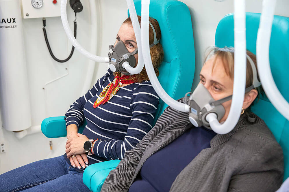
In hyperbaric environment (usually at 2-3 atmospheres pressure), blood can dissolve much more oxygen than normally. Practically, plasma becomes very rich in oxygen, which thus reaches tissues much more efficiently, even where circulation is deficient. Additionally, increased pressure has direct effect of edema reduction (decreases volume of excess gases and liquids, according to Boyle's law) and decreases inflammation and pain. During a typical hyperbaric oxygen therapy session, patient sits in sealed chamber, breathing 100% oxygen for approximately 120 minutes, at 2-2.5 atmospheres pressure, with decompression breaks included.
For lumbar bone edema, hyperbaric oxygen therapy has several theoretical and practical benefits:
- Increased bone oxygenation: Bone normally has reduced vascularization compared to other organs, so bringing oxygen surplus helps bone cells (osteoblasts, osteoclasts) and bone marrow cells function optimally. Oxygen stimulates local metabolism and healing processes – favors new bone tissue formation and affected area resorption. In vivo study demonstrated that hyperbaric therapy stimulates HIF-1 growth factor production (hypoxia-inducible factor 1), which in turn triggers tissue repair cascade.
- Angiogenesis (new vessel formation): Bone tissue affected by edema often suffers from inadequate perfusion. Hyperbaric therapy "forces" oxygen to diffuse deep into tissues and encourages new blood vessel formation in injured area. The sooner new capillaries appear, the faster normal nutrient and oxygen supply to bone recovers, hastening healing.
- Inflammation and edema reduction: Hyperbaric environment has anti-inflammatory effect; decreased levels of inflammatory mediators such as TNF-α and interleukin-6 have been observed in patients treated with hyperbaric oxygen for osteonecrosis. Also, hyperbaric oxygen combats excessive oxidative stress through free radical modulation (paradoxically, although providing extra oxygen, long-term reduces oxidative inflammation). Many people also report pain reduction after several sessions, possibly through decreased interstitial pressure in bone (edema shrinks) and direct effect on nociceptive receptors.
- Clinically demonstrated efficiency: Although relatively newly introduced therapy for bone edema, there are studies and case reports supporting its efficacy. Study published in Medicine (Baltimore) journal in 2023 concluded that hyperbaric oxygen therapy is effective treatment option for bone marrow edema, determining faster healing compared to lack of this treatment and without significant adverse effects. Also, for knee bone edemas (medullary edema syndrome) excellent results have been obtained: in retrospective study, 81–88% of patients with incipient osteonecrosis treated with hyperbaric oxygen presented complete MRI healing, compared to only 17% in control group without HBOT. At ~11 year follow-up, 93% of those patients maintained joint without needing prosthesis, highlighting therapy's lasting effect. In another comparative study on patients with knee bone edema from degenerative causes, 35% reduction in edema area on MRI was found in group treated with aspirin + bisphosphonates + hyperbaric therapy, versus only 5% reduction in those treated only with medications, without hyperbaric oxygen therapy. Practically, adding hyperbaric therapy significantly accelerated edema resorption. These results from knee and hip orthopedics are encouraging and can be extrapolated to lumbar spine level, where bone healing mechanisms are similar.
Hyperbaric oxygen therapy is becoming more accessible in Romania too, but it's essential that this treatment be performed in accredited center, respecting highest safety standards. Hyperbarium clinic is among few units specialized in hyperbaric medicine, offering patients not only one of most modern hyperbaric chambers in country, but also medical staff with specific training and rigorous evaluation before, during, and after treatment completion. Choosing such center is essential for therapy efficiency and safety.
Additionally, at Hyperbarium clinic emphasis is placed on multidisciplinary approach: hyperbaric therapy is integrated with other necessary treatments (physiotherapy, medication, orthopedic or rheumatological consultation), so patient has complete treatment plan for lumbar bone edema. Practically, hyperbaric therapy doesn't replace other therapies, but potentiates them. For example, patient can follow parallel kinesiotherapy and hyperbaric oxygen sessions, thus obtaining both mechanical and biological benefits in bone healing.
Conclusions and advice for patients
Although most lumbar bone edema cases are treated conservatively, in approximately 10–15% of situations interventional treatment is necessary, especially when pain persists or there's vertebral collapse risk. In Romania, osteoporotic vertebral fracture incidence is increasing, affecting over 25,000 people annually, especially women over 65 years old.
Thus, lumbar bone edema may sound alarming, but in reality it's warning signal, not condemnation. With correct and timely treatment, most patients recover completely. Important is identifying cause: can be trauma, discopathy, or metabolic problem, such as osteoporosis, condition affecting over 1 million Romanians, mostly postmenopausal women. Treatment varies from rest and kinesiotherapy to modern procedures like hyperbaric oxygen or vertebroplasty.
In Romania, back pain is one of most frequent causes of medical leave, and lack of timely intervention can worsen situation.
Listen to your body, follow medical plan, attend physiotherapy sessions, and don't hesitate to ask for second opinion. With support of right medical team, such as that at Hyperbarium, and with perseverance, you can return to active life free of pain.