


Article reviewed by: Dr. Sturz Ciprian, Dr. Tîlvescu Cătălin and Dr. Alina Vasile
Necrosis of the femoral head - What it is, Causes, Symptoms and Treatment
- What is femoral head necrosis?
- How do we recognize the symptoms in femoral head necrosis?
- What causes femoral head necrosis?
- How is femoral head necrosis diagnosed?
- Misdiagnosis - a real, global problem
- What are the treatments for femoral head necrosis?
- Revolutionary technique for treating necrosis
- How does hyperbaric therapy work in bone necrosis?
- Effects of hyperbaric therapy in femoral head necrosis
- Experience of the Hyperbarium clinic in the treatment of bone necrosis
- Complications that can occur with femoral head necrosis
- Methods to prevent femoral head necrosis
What is femoral head necrosis?
Bone necrosis, also known as osteonecrosis or avascular necrosis, is a condition in which part of the bone dies due to interruption of blood flow. Without adequate blood supply, the bone tissue begins to break down and over time the bone may collapse.
This condition can occur in any bone, but is most commonly found in the femoral head (the upper part of the thigh bone), affecting the hip joint. If not treated in time, avascular necrosis evolves into coxarthrosis, a condition that is manifested by the progressive deterioration of the hip joint.
In this article, we will explore not only the causes and symptoms of femoral head necrosis, but also the main diagnostic methods, along with the most modern and effective treatments available for this condition.
How do we recognize the symptoms in femoral head necrosis?
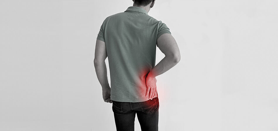
Necrosis of the femoral head is manifested by a series of symptoms that worsen as the condition progresses. In the early stages, symptoms may be subtle or absent, but as the disease progresses, signs and symptoms become more apparent. This condition occurs more frequently in people between the ages of 35 and 50, affecting especially men, with the ratio being about eight men for every woman affected.
Main manifestations include:
- Pain: Initially, the pain may be mild and intermittent, felt in the groin, buttocks or front of the thigh. As the condition progresses, the pain becomes constant and intense, increasing during physical activities and decreasing at rest. The pain can also be felt during the night or when changing the position of the body.
- Limitation of movements: Mobility of the hip joint becomes reduced, especially for movements such as internal and external rotation of the hip. Patients may have difficulty getting up from a chair, climbing stairs, or performing other daily activities.
- Lamping: Due to pain and loss of joint function, the patient may begin to limp.
- Feeling unstable: The hip joint may appear unstable or loose, increasing the risk of falls and injury.
- Muscle atrophy: The muscles around the hip and thigh can begin to atrophy due to lack of use and constant pain.
- Shortening of the affected limb: In advanced cases, the collapse of the femoral head can lead to the shortening of the affected leg.
All these symptoms worsen as the disease progresses, making early diagnosis essential to stop its progression and reduce the risk of complications.
What causes femoral head necrosis?
Necrosis of the femoral head, or avascular osteonecrosis, is caused by interruption of the blood supply to the femoral head, leading to bone cell death and structural collapse of the bone.
This disruption can have various causes and risk factors, which include:
- Trauma: Fractures or dislocations of the hip can affect blood flow to the femoral head.
- Chronic use of corticosteroids: Long-term use of corticosteroids, used to treat conditions such as asthma, rheumatoid arthritis and other inflammatory diseases, is a major risk factor.
- Excessive alcohol consumption: Excess alcohol can damage blood vessels and cause fat to build up in the blood, reducing blood flow to the bone.
- Medical conditions: Conditions such as systemic lupus erythematosus, diabetes, HIV/AIDS, inflammatory bowel disease, and other autoimmune diseases can increase the risk of osteonecrosis.
- Radiotherapy treatment: Radiation therapy used in the treatment of cancer can damage blood vessels and bone structure, leading to necrosis.
- Blood clotting disorders: Conditions that affect blood clotting, such as thrombophilia, can affect blood flow to the bone.
- Hypertension: High blood pressure can damage the small blood vessels that supply the femoral head.
- Metabolic and endocrine diseases: Conditions such as Gaucher disease, hyperlipidemia and other metabolic disorders can contribute to the development of necrosis of the femoral head.
- Smoking: Nicotine and other chemicals in cigarettes can affect blood circulation and bone health.
- Dysfunctions of normal biomechanics: These can lead to necrosis of the femoral head through uneven weight distribution, increased joint pressure, repetitive stress and limitation of mobility, thus affecting blood circulation and bone health.
In some cases, even after detailed investigations, the cause of femoral head necrosis may remain unidentified and is classified as idiopathic. This means there are no known genetic factors, pre-existing diseases or exposures to explain the disease.
How is femoral head necrosis diagnosed?
Diagnosing femoral head necrosis involves a combination of history, physical examination, and imaging investigations. Here are the typical steps for diagnosis:
- Anamnesis:
- Medical history: The doctor will ask about symptoms, onset and duration of pain, and any known risk factors (eg, corticosteroid use, alcohol consumption, trauma).
- Symptoms: Details related to the location and nature of pain, changes in mobility, and activities that worsen or relieve pain will be discussed.
- Physical examination:
- Palpation and motion assessment: The doctor will examine the hip for tenderness, assess range of motion, and look for signs of muscle atrophy or differences in leg length.
- Functional tests: Tests will be performed to assess the stability of the hip and the patient's ability to move.
- Imaging investigations:
- X-ray: In early stages, x-rays may appear normal, but in more advanced stages they may show collapse of the femoral head and changes in bone structure.
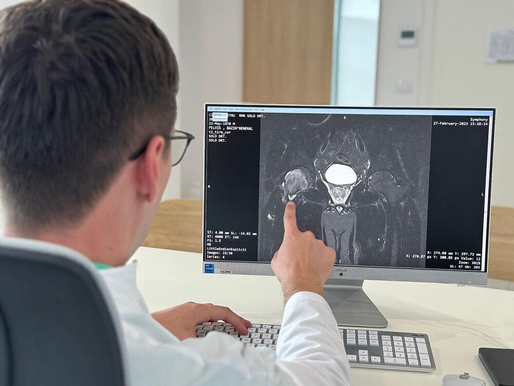
- Nuclear Magnetic Resonance (MRI): It is the most sensitive and specific method for detecting necrosis of the femoral head, especially in the early stages. MRI can reveal changes in bone structure and blood supply.
- Computed tomography (CT): CT can provide additional details about the bone structure and the degree of collapse of the femoral head.
- Bone scintigraphy: This can be used to assess bone metabolism and detect areas of reduced blood supply.
- Lab tests:
- Although not specific for the diagnosis of necrosis of the femoral head, certain blood tests may be performed to evaluate possible underlying causes, such as coagulation disorders, lipid levels, or inflammatory markers.
- Bone biopsy:
- Rarely, in cases where the diagnosis is unclear, a bone biopsy may be performed to confirm the presence of necrosis and to rule out other conditions.
Misdiagnosis - a real, global problem
Until recently, avascular necrosis was thought to be responsible for approximately 10% to 12% of all hip replacements performed globally. Other major causes include osteoarthritis, rheumatoid arthritis, and trauma.
However, new studies (study 1, study 2) shows that avascular necrosis can often be misdiagnosed in the early stages due to nonspecific initial symptoms . In some cases, misdiagnosis rates can vary between 20% and 40%, depending on the diagnostic methods used and the level of expertise of the doctors.
Only in Romania, the reported number of hip replacement operations is over 10,000 every year, which indicates that approximately 500 patients annually do not receive a correct diagnosis in time, significantly contributing to the high number of operations and emphasizing the importance of correctly identifying the condition in its early stages.
Patients who suspect they may have avascular necrosis or are concerned about a possible misdiagnosis can take several steps to increase their chances of getting a correct diagnosis:
- Consult a specialist: Requesting a consultation with an orthopedic or rheumatologist specializing in musculoskeletal disorders can help to identify the correct diagnosis.
- Advanced imaging investigations: Order an MRI (nuclear magnetic resonance). Standard radiographs or CT (computed tomography) scans may not be sufficient to diagnose the early stages of bone necrosis.
- Get a second opinion: If the initial diagnosis is unsatisfactory or unclear, getting a second opinion from another specialist or hospital can provide additional insight and increase the chances of a correct diagnosis.
- Communicate medical history: Clear communication of medical history (such as history of trauma, alcohol consumption, or steroid use) and risk factors can help the doctor recognize avascular necrosis more easily.
- Monitor symptoms: Note changes in symptoms and discuss them with your doctor.
For an initial consultation or a second opinion, patients can confidently turn to the doctors of the Hyperbarium clinic, where they will benefit from a personalized, complex evaluation and treatment, under the careful monitoring of specialized medical staff.
What are the treatments for femoral head necrosis?
Until recently, non-surgical treatment of bone necrosis included medications to reduce pain and inflammation, physiotherapy, activity modification to reduce pressure on the affected joint, and the use of braces. Unfortunately, these options cannot completely stop the progression of necrosis and avoid hip replacement surgery (arthroplasty).
One of the most commonly used surgical options for treatment is bone decompression, which involves drilling holes in the affected bone to release internal pressure and stimulate the formation of new blood vessels, helping to regenerate it.
Another surgical option that aims to avoid or delay hip replacement surgery is the addition of bone grafts, to support the structure and try to promote bone regeneration, especially in the early stages of necrosis bones.
Other therapies still in the experimental or unapproved phases, but which may be widely used in the future, include stem cell therapy, gene therapy and nanotherapy.
Revolutionary technique for treating necrosis
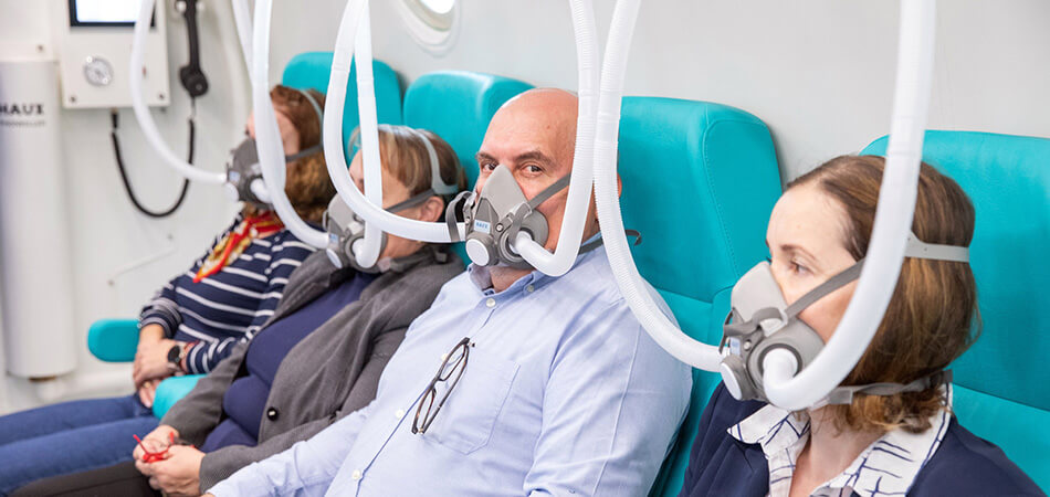
A revolutionary solution, increasingly recognized in the medical community for treating necrosis of the femoral head, is hyperbaric therapy. The success rate of this therapy, especially in the early stages, is over 80%, in many cases total healing is achieved.
By exposing the patient to pure oxygen in a special chamber, pressurized above atmospheric pressure, this non-invasive and painless therapy stimulates the formation of new blood vessels and promotes the regeneration of damaged bone, providing results remarkable.
After numerous studies have shown that hyperbaric oxygen therapy (HBOT) is an effective solution for treating necrosis of the femoral head, especially in the early stages (grade I and II), in 2016, at the Hyperbaric Medicine Conference in Lille, France, the European medical community included femoral head necrosis among the indications for hyperbaric oxygen therapy.
How does hyperbaric therapy work in bone necrosis?
Under hyperbaric pressure conditions of 3 ATA (absolute pressure) and administration of 100% pure oxygen, blood oxygen and hemoglobin levels are increased up to 20 times, and oxygen can be diffused to a depth of up to 3 times higher in tissues than normal. Thus, hyperbaric oxygen therapy significantly contributes to the diffusion of oxygen in poorly oxygenated tissues, favoring cellular metabolism and accelerated healing.
Effects of hyperbaric therapy in femoral head necrosis
Hyperbaric oxygen therapy (HBOT), through its hyperoxic effect (exposure to high levels of oxygen), produces various physiological effects that are beneficial in the treatment of femoral head necrosis, as follows:
- Vascularizes bone tissue: by increasing the amount of oxygen in the tissues and by stimulating growth factors such as vascular endothelial growth factor (VEGF), hyperbaric therapy stimulates the formation of new blood vessels in bone tissue.
- Reduces inflammation and pain: hyperbaric therapy reduces inflammatory markers and improves blood circulation, leading to decreased bone edema and pain.
- Stimulates bone regeneration: hyperbaric oxygen therapy stimulates the production of osteoblasts, specialized cells derived from stem cells, responsible for the formation of bone tissue.
- Stimulates collagen synthesis: activates fibroblasts to synthesize collagen fibers, an essential protein for the strength of bones and joints.
- Reduces oxidative stress: stimulates the release of detoxification enzymes and antioxidant molecules, which help neutralize free radicals and maintain a healthy redox balance in the body.
- Prevents bone infections: hyperbaric therapy increases immunity by stimulating the activity of leukocytes, white blood cells that fight infections and other types of tissue damage.
To provide as many answers as possible to both patients and doctors, a collection of scientific studies confirming the effectiveness of hyperbaric therapy in the treatment of bone necrosis has been published on the website of the Hyperbarium clinic.
Now, more and more doctors are recommending this therapy to treat necrosis in the early stages or to stop the progression of the disease in more advanced stages and to prevent the onset of coxarthrosis (arthrosis of the hip).
Experience of the Hyperbarium clinic in the treatment of bone necrosis
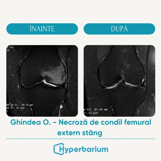
At the Hyperbarium clinic, our doctors have documented numerous cases of bone necrosis successfully treated with hyperbaric therapy. An example is the case of a 54-year-old patient, who was diagnosed by MRI with necrosis of the left external femoral condyle.
The initial treatment plan included a cycle of 20 hyperbaric therapy sessions, which reduced pain intensity by 60% and reduced edema by 80%.
After completing the 40 treatment sessions, the patient's pain decreased from 10/10 to 2/10 and the edema reduced by 95%, highlighting the effectiveness of this therapy and providing a very good prognosis. The patient did not follow any other treatments or therapies during this time.
Although aseptic necrosis of the femoral head is the most common type of necrosis treated by hyperbaric therapy, bone necrosis can occur in any bone: knee (femoral and tibial condyle ), shoulder (humeral head), ankle (talus), but also small bones of the hand and foot.
To fully understand hyperbaric therapy and its effectiveness in the treatment of necrosis of the femoral head, we invite you to read this complete guide. You will find answers to all questions, details about the whole process, but also information about the price.
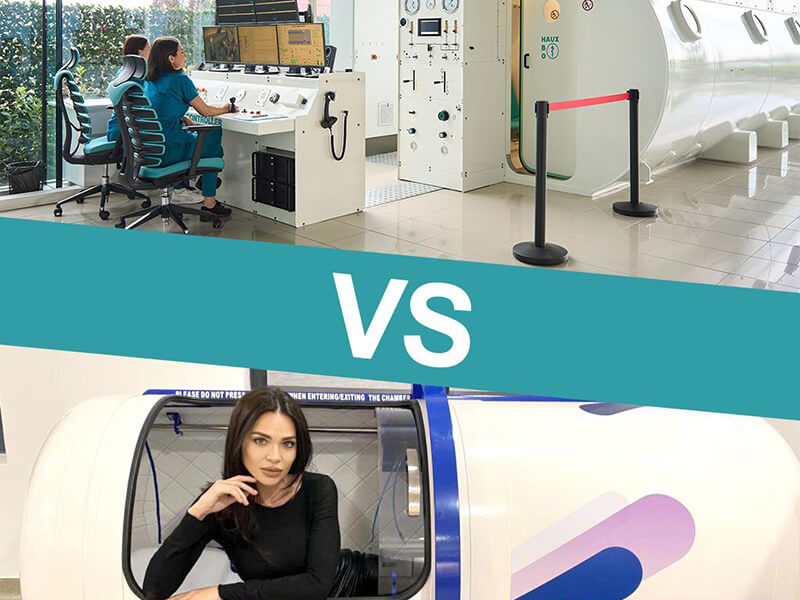
The Hyperbarium Clinic is one of the most advanced and modern hyperbaric medicine centers in Romania. The entire staff is trained and certified according to European standards, and the team doctors have skills in hyperbaric medicine. The medical director of the clinic is Dr. Ciprian Sturz, a surgeon specializing in general surgery, visceral and emergency surgery, with 15 years of experience in Germany.
If you have symptoms of femoral head necrosis or persistent hip pain, we encourage you to schedule a consultation to explore treatment options that can not only improve significantly the quality of life, but they can also achieve a total cure.
Complications that can occur in femoral head necrosis
Femoral head necrosis can lead to a number of serious complications if not treated in time. These complications result from progressive damage to the hip bone and joint. Here are the main complications that can occur:
- Femoral head collapse: This is a serious complication of necrosis, causing major damage to the upper part of the thigh bone that forms the hip joint. It occurs when the necrotic bone, weakened by the lack of blood supply, can no longer bear the weight and normal stress, leading to its collapse.
- Secondary degenerative arthritis: Damage and deformation of the femoral head can lead to the development of degenerative arthritis (osteoarthritis) in the hip joint. This can cause chronic pain and severe limitation of movements.
- Leg length discrepancy: Femoral head collapse can lead to shortening of the affected limb, resulting in a leg length discrepancy. This can cause additional walking problems and pain in other parts of the body, such as the back or knee.
- Chronic pain: Uncorrected femoral head necrosis can lead to severe and constant pain, affecting quality of life and ability to perform daily activities.
- Reduced Mobility: As the hip joint deteriorates, mobility can be severely limited. Patients may have difficulty moving, standing, or carrying out normal daily activities.
- Deformation of the joint: Structural changes in the femoral head and the acetabulum (the cavity in which the femoral head articulates) can lead to the deformation of the hip joint, affecting its function.
- Bilateral necrosis: In some cases, the necrosis may also affect the other hip, further exacerbating mobility difficulties and pain.
- Secondary osteoporosis: Prolonged inactivity and immobilization due to hip pain and dysfunction can lead to secondary osteoporosis, which increases the risk of fractures in other areas of the body.
- Need for surgery: In advanced stages, necrosis of the femoral head may require major surgery, such as osteotomy (reshaping the bone) or total hip arthroplasty (replacing the hip joint). If surgery is necessary, there is a risk of postoperative infection, which can complicate the recovery process and require additional antibiotic treatment or even reoperation.
- Psychological problems: Chronic pain and functional limitation can lead to mental health problems such as depression and anxiety. The psychological impact of the inability to carry out daily activities and participate in social life can be significant.
To prevent these complications, early diagnosis and appropriate treatment of femoral head necrosis is essential. In stages 1 and 2, hyperbaric therapy can lead to complete healing in over 80% of cases.
Methods to prevent femoral head necrosis
Preventing necrosis of the femoral head involves several aspects of lifestyle and health care to avoid risk factors that can lead to this condition.
First, it is important to take care of your general health. If you need to take corticosteroids (strong anti-inflammatory drugs) for a long time, please talk to your doctor about the risks and possible alternatives. These drugs can reduce blood flow to the bones, so they should be used with caution.
Alcohol consumption must be limited. Excess alcohol can cause fat to build up in the blood vessels, which can block blood flow to the femoral head. So, maintaining moderate alcohol consumption or avoiding it completely is essential.
If you smoke, quitting is crucial. Smoking affects blood circulation and can contribute to damage to the vessels that supply bones. Quitting smoking improves cardiovascular health and reduces the risk of many diseases, including necrosis of the femoral head.
Avoiding hip injuries is also important. While you can't prevent all accidents, wearing the right protective gear during sports or work and practicing good lifting technique can help reduce the risk of injury.
Maintaining a balanced diet and an active lifestyle contributes to bone and joint health. Make sure your diet includes enough calcium and vitamin D, which are essential for bone health. Regular exercise, such as walking, swimming or cycling, helps maintain bone density and muscle strength, which can prevent joint damage.
If you suffer from a medical condition that may predispose to femoral head necrosis, such as lupus or diabetes, it is essential that you follow the prescribed treatment and go for regular medical check-ups. Careful monitoring and proper management of these conditions can reduce the risk of complications.
In short, preventing necrosis of the femoral head involves a healthy lifestyle, limiting alcohol consumption and quitting smoking, avoiding trauma, maintaining a balanced diet and regular exercise, and properly managing pre-existing medical conditions. All these measures contribute to maintaining good blood circulation to the bones and preventing their damage.
Modern treatments, such as hyperbaric therapy, bring a ray of hope to patients, because in the early stages of necrosis, this method leads to complete healing in most cases, demonstrating the significant evolution and considerable progress made in the medical field in the last decade.