


Article reviewed by: Dr. Sturz Ciprian, Dr. Tîlvescu Cătălin and Dr. Alina Vasile
Article updated on: 08-08-2026
Hyperbaric therapy and cancer: what HBOT can and cannot do
- What is the connection between oncology and hyperbaric therapy?
- Cancer is not a single disease
- Radiotherapy: what it means and how it works
- Acute and delayed effects of radiotherapy
- What is hyperbaric therapy?
- What hyperbaric oxygen cannot do in oncology
- How hyperbaric oxygen helps irradiated tissue
- Can you undergo hyperbaric therapy if you have untreated cancer or are receiving treatment?
- How hyperbaric therapy is integrated into cancer treatment
- How hyperbaric therapy is performed at Hyperbarium
- What to do after radiotherapy
- When should you seek medical evaluation quickly?
- The real place of hyperbaric therapy in oncology
Many patients raise the question of how much their body has been affected after completing radiotherapy sessions. Radiotherapy behaves a little like a controlled storm. It knows exactly where to strike to destroy the tumor, but a few drops inevitably reach the neighboring healthy tissue as well. In most patients, this tissue recovers on its own, like grass after rain. In some, however, the “soil” remains too poor in oxygen to regenerate by itself, and this is where hyperbaric therapy has a role. It is not a cancer treatment, but a supportive treatment for recovery.
This article is not about curing cancer with oxygen under pressure, because HBOT does not do that, and anyone who claims otherwise is distorting the medical literature. Instead, it discusses a much more concrete and scientifically better-documented area: recovery after a course of radiotherapy.
What is the connection between oncology and hyperbaric therapy?
The question of what follows radiotherapy does not have the same answer for every patient. For some, it is followed by periodic checkups and recovery without major problems. For others, cancer treatment continues through surgery, chemotherapy, immunotherapy, or hormone therapy. There are also patients who develop injuries to healthy tissues exposed to radiation months or even years later. In this last situation, hyperbaric oxygen therapy, also known by the abbreviation HBOT, may become relevant.
It is essential to establish a clear boundary from the outset: hyperbaric therapy does not treat cancer directly. It does not destroy the tumor, replace surgery, chemotherapy, radiotherapy, immunotherapy, or the treatment prescribed by the oncologist, and it must not be presented as an alternative method of curing cancer.
In oncology, the best-established medical role of HBOT is the treatment of certain injuries caused by radiotherapy to healthy tissues. These include problems such as mandibular osteoradionecrosis, chronic soft-tissue injuries, radiation cystitis, and, in carefully selected situations, radiation proctitis. These are complications of cancer treatment, not tumors treated with oxygen.
This distinction is important both medically and humanly. A patient who has gone through cancer does not need spectacular promises, but precise explanations: what problem the physicians are trying to treat, what evidence exists, what limitations the therapy has, and how it fits into the plan established by the oncology team.
Cancer is not a single disease
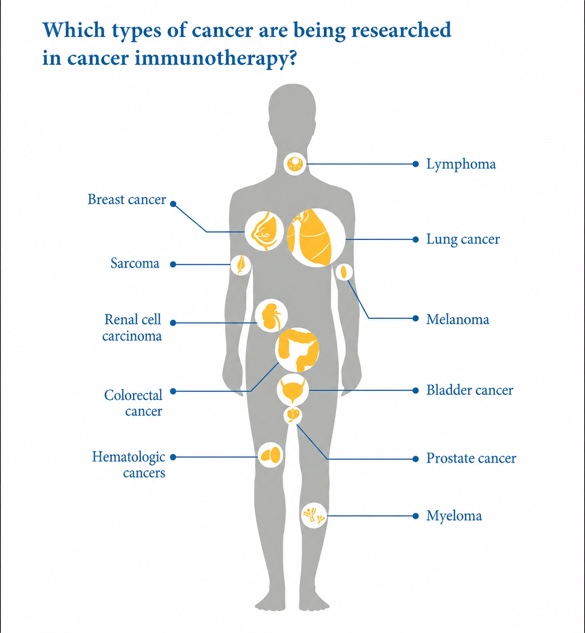
We use the word “cancer” as an umbrella term for many conditions that share a common feature to some extent: the appearance of altered cells that multiply uncontrollably, may invade surrounding tissues, and, in some cases, can spread to other regions of the body through metastases.
The main categories of cancer, classified according to the tissue from which they arise, are:
- Carcinomas—the most common types of cancer—begin in epithelial cells that cover the skin, organs, and body cavities; this category includes many cancers of the breast, lung, prostate, colon, bladder, cervix, head, and neck
- Sarcomas develop from tissues such as bone, muscle, fat, or blood vessels
- Leukemias begin in blood-forming tissues, usually the bone marrow, and do not always produce a solid tumor
- Lymphomas affect the lymphatic system
- Multiple myeloma begins in plasma cells, which are cells of the immune system
- Other categories include tumors of the central nervous system, germ-cell tumors, and numerous rare forms, each with its own biological behavior
Therefore, treatment is not chosen only according to the organ in which the tumor was found. The cell type, stage of disease, molecular profile, local extent, presence of metastases, general health, and the patient’s other illnesses all matter.
Treatment may include:
- surgery, which removes the tumor;
- chemotherapy, which uses medicines that act especially on rapidly dividing cells;
- radiotherapy, which damages the DNA of tumor cells;
- immunotherapy, which helps the immune system recognize and attack cancer;
- targeted therapy, directed at specific proteins or molecular changes;
- hormone therapy, used for cancers influenced by hormones, such as certain breast or prostate tumors.
In many cases, these methods are combined.
In Romania, cancer is one of the leading causes of death. According to the Country Cancer Profile published by the OECD and the European Commission in 2025, 45,866 cancer deaths were recorded in 2022, while neoplasms were the second leading cause of death after cardiovascular diseases, accounting for approximately 19% of all deaths in 2019. Worldwide, the World Health Organization and the International Agency for Research on Cancer estimated approximately 20 million new cases in 2022, and with population aging and exposure to risk factors, the number could exceed 35 million by 2050; the WHO details this growth in the global cancer burden. These figures explain why modern medicine focuses not only on curing or controlling the tumor, but also on the patient’s life after treatment.
Radiotherapy: what it means and how it works
Radiotherapy is a treatment that uses high doses of ionizing radiation to damage the DNA of tumor cells. When the DNA damage is extensive enough, cancer cells can no longer divide and die. The effect is not always immediate. The process may continue for weeks or months after the last session. The National Cancer Institute explains the mechanism of radiotherapy and the differences between external-beam radiation, brachytherapy, and systemic radionuclide treatments.
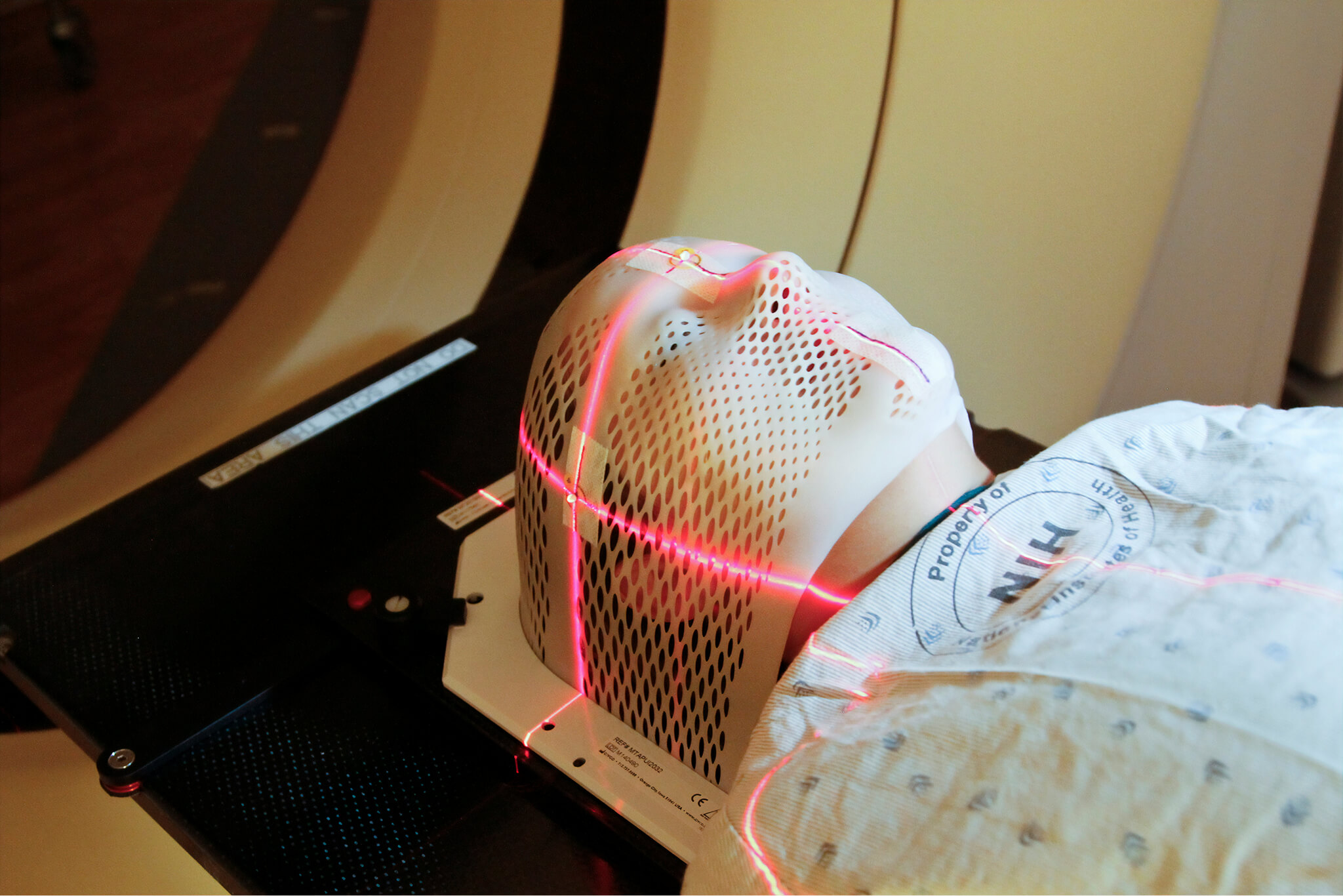
Radiotherapy is a local treatment. For example, if the prostate is irradiated, the main effect is sought in the pelvis; if a tumor in the head and neck region is treated, the beams are concentrated on that region. Modern planning uses imaging, computer systems, and techniques such as IMRT, which modulates beam intensity to distribute the dose as precisely as possible and protect neighboring organs. Nevertheless, healthy tissue inevitably lies between the machine and the tumor. Medical physics can reduce the dose received by that tissue, but it cannot eliminate it in every situation, and radiation may affect normal cells, small blood vessels, connective tissue, and the local capacity for repair.
Radiotherapy is used to cure certain cancers, reduce the risk of recurrence after surgery, shrink the tumor before an operation, or control symptoms in advanced disease. The International Atomic Energy Agency estimates that approximately 50–70% of cancer patients need radiotherapy at some point. Consequently, people who undergo radiotherapy are a very large and diverse group: some receive only a few sessions, others follow a protocol lasting several weeks, and both the dose and the treated area vary considerably.
Acute and delayed effects of radiotherapy
Not every reaction that appears after irradiation is a late complication, and not every reaction is an indication for HBOT. Most radiotherapy side effects are acute: they occur during treatment or in the first weeks afterward and are related to inflammation of the exposed tissues.
Depending on the region treated, patients may experience skin redness and sensitivity, fatigue, hair loss only within the irradiated field, inflammation of the oral mucosa, difficulty swallowing, nausea, diarrhea, frequent urination, or pelvic discomfort. Many of these reactions diminish after radiotherapy ends as healthy cells recover.
Delayed injuries are different. They may appear after six months, several years, or sometimes more than a decade. Instead of temporary inflammation, there may be a slow deterioration of the microcirculation: small vessels narrow, tissue becomes fibrotic, less elastic, poorly vascularized, and chronically low in oxygen. Three terms are sometimes used in hyperbaric medicine to describe irradiated tissue: hypoxic, meaning it receives too little oxygen; hypovascular, meaning it has too few functioning blood vessels; and hypocellular, indicating that the number and activity of cells capable of repairing the area are reduced.
This is a difficult cycle to break. Tissue repair requires oxygen and circulation, but radiation has damaged the circulation itself. A minor wound, a dental extraction, an infection, or later surgery may exceed the region’s capacity to heal. These effects do not mean that radiotherapy was performed incorrectly; in many situations it was necessary to control the cancer and saved the patient’s life. Late injury is a possible consequence of exposing healthy tissues to the dose required to treat the tumor.
What is hyperbaric therapy?
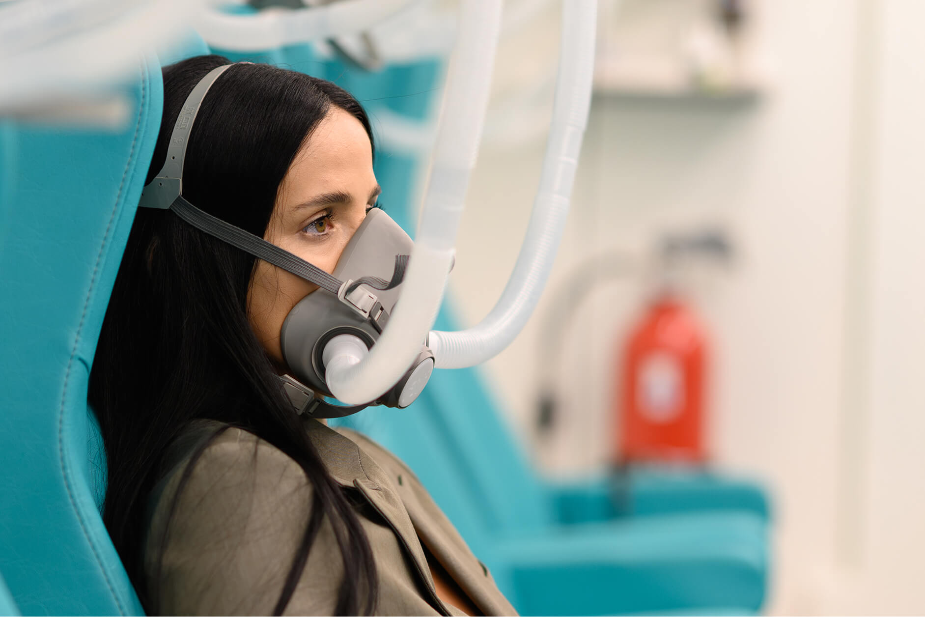
Hyperbaric oxygen therapy requires the patient to breathe medical oxygen at a concentration close to 100% inside a chamber where the pressure is higher than normal atmospheric pressure. At sea level, we breathe air containing approximately 21% oxygen at a pressure of 1 ATA. During HBOT, treatment pressure is frequently around 2–2.5 ATA. Pressure is not an unimportant technical detail: it changes the way oxygen enters the blood and is distributed throughout the tissues.
Under normal conditions, most oxygen is carried by hemoglobin in red blood cells. Under pressure, the amount of oxygen dissolved directly in blood plasma increases significantly, allowing the plasma to carry oxygen toward areas where microcirculation is impaired, although this effect cannot completely replace destroyed blood vessels.
For a more detailed explanation of the physical and biological mechanism, see Hyperbarium’s article on how hyperbaric therapy works.
During a session, pressure is increased gradually. The patient may feel a sensation in the ears similar to takeoff or landing in an airplane. Once the working pressure is reached, the oxygen exposure period begins, sometimes divided by air breaks, and the chamber is slowly depressurized at the end. For late post-radiation injuries, protocols used in medical practice often involve repeated sessions, usually on consecutive weekdays for several weeks.
What hyperbaric oxygen cannot do in oncology
Searching for the phrase “hyperbaric therapy cancer” can produce very different results: laboratory studies, hypotheses about tumor oxygenation, commercial advertisements, personal testimonials, and information about post-radiotherapy injuries. These categories must not be confused.
Hyperbaric therapy is not a stand-alone cancer treatment. There is insufficient clinical evidence to claim that a standard course of hyperbaric oxygen therapy cures a tumor, predictably shrinks it, eliminates metastases, or prevents recurrence. The therapy does not replace biopsy, histopathological diagnosis, staging, surgery, chemotherapy, radiotherapy, immunotherapy, or targeted treatment and must not be used to delay the recommended cancer treatment.
The distinction between complementary and alternative therapy is equally important. Complementary cancer therapies are used alongside standard treatment for a clear indication and after risks have been assessed. An alternative therapy is used instead of proven cancer treatment, and this substitution may reduce the chances of survival. The National Cancer Institute explicitly emphasizes this distinction and explains that integrative medicine must combine standard treatment with methods for which there is reasonable evidence of safety and usefulness in its overview of complementary and alternative medicine. In the context discussed here, HBOT is an adjunctive medical treatment for certain consequences of radiotherapy. The target is damaged healthy tissue, not the tumor.
How hyperbaric oxygen helps irradiated tissue
Healthy tissue heals through a sequence of coordinated processes. Immune cells clean the area, fibroblasts produce collagen, endothelial cells contribute to blood-vessel formation, and the tissue receives nutrients and oxygen through local circulation. After irradiation, this mechanism may be slowed or blocked: small vessels become scarce and fragile, collagen is deposited abnormally, fibrosis makes the tissue rigid, and repair cells no longer function efficiently in an oxygen-poor environment.
HBOT temporarily increases the partial pressure of oxygen in tissue. Controlled repetition of these increases can activate biological signals involved in angiogenesis—the formation of new blood vessels—and in recruiting cells required for repair. This is not about permanently “filling” the tissue with oxygen after a single session; the intended effect appears through repetition of a stimulus. Oxygenation rises considerably inside the chamber and gradually returns toward its initial level afterward, while the repeated alternation may support the cellular response and microvascular reconstruction.
The European Consensus Conference on Hyperbaric Medicine and the Undersea and Hyperbaric Medical Society include late radiation injuries among the recognized clinical uses of HBOT. Nevertheless, a plausible mechanism does not guarantee success for every patient. The result depends on the type of injury, degree of fibrosis, presence of infection, vascular health, smoking, diabetes, associated treatments, and any surgical procedures. Some injuries respond well, others only partially, and surgery may remain necessary in advanced cases.
Mandibular osteoradionecrosis and hyperbaric therapy
Mandibular osteoradionecrosis occurs when irradiated bone loses its vitality and can no longer heal normally. The best-known form affects the mandible after radiotherapy for head and neck cancers. The mandible has a poorer blood supply than other facial bones and is subjected every day to the pressure of chewing, so a dental extraction, a denture that injures the gum, or an infection can trigger bone exposure and necrosis in an irradiated patient.
The patient may notice persistent pain, visible bone in the mouth, wounds that do not close, discharge, unpleasant breath, recurrent infections, difficulty opening the mouth, chewing, or swallowing, and in advanced stages fistulas and pathological fractures may occur. In this complication, hyperbaric therapy must not be viewed in isolation. Treatment may include oral hygiene measures, antibiotics when infection is present, pain control, removal of necrotic tissue, and, in severe cases, surgical resection and reconstruction. Hyperbaric oxygen therapy may be used before and after surgery to prepare the tissue bed and support healing.
The European consensus recommends hyperbaric oxygen therapy for mandibular osteoradionecrosis, but contemporary medical literature requires an important qualification: evidence about treating an established lesion must not be confused with routine prevention before every dental extraction. The randomized HOPON study, published in 2019, examined prevention of osteoradionecrosis in patients with an irradiated mandible who were due to undergo dentoalveolar procedures. The incidence at six months was 6.4% in the HBOT group and 5.7% in the control group, with no significant difference. The result shows that hyperbaric prophylaxis should not be applied automatically to every patient who has received radiotherapy, but should be decided individually according to the dose received by the mandible, the condition of the bone, the type of procedure, and other risk factors.
Radiation cystitis and HBOT
Radiation cystitis is chronic inflammation of the urinary bladder caused by pelvic radiotherapy and can lead to bleeding, pain, and frequent, painful urination. The five-year follow-up of the RICH-ART study showed that improvement was maintained for many participants, with an average improvement of 19.1 points in the urinary score compared with baseline—above the threshold considered clinically important for that instrument. This is a valuable result because radiation cystitis is a chronic condition, and the goal is not merely an improvement lasting a few weeks.
HBOT is not, however, the first response to every episode of blood in the urine. Heavy hematuria, retention caused by clots, dizziness, weakness, or a drop in hemoglobin may require urgent urological treatment rather than an appointment for hyperbaric therapy.
How hyperbaric therapy helps radiation proctitis and intestinal injuries
Radiation proctitis is injury to the rectum following pelvic radiotherapy. It may cause bleeding, mucus, an urgent need to defecate, diarrhea, pain, and tenesmus—the persistent feeling that the rectum has not emptied—and, in severe cases, ulceration, stenosis, or fistulas. Treatment may include dietary changes, local medicines, sucralfate, endoscopic procedures such as argon plasma coagulation, and surgery in complicated cases. HBOT is one possible option for chronic forms that are refractory to usual treatment, but the evidence is less consistent than for radiation cystitis.
A double-blind randomized study published in 2008 evaluated 120 eligible patients with refractory radiation proctitis. The proportion considered responders was 88.9% in the hyperbaric group versus 62.5% in the control group, and the researchers calculated a 32% absolute reduction in the risk of failure, equivalent to treating approximately three patients to obtain one additional response; the study can be consulted on PubMed. The 2023 Cochrane review concludes that hyperbaric therapy may improve certain late tissue injuries in the head, neck, and pelvis and may reduce the risk of wound breakdown in irradiated tissues, but the authors emphasize that many studies included few patients, different methods, and varying levels of quality.

Can you undergo hyperbaric therapy if you have untreated cancer or are receiving treatment?
The short answer is complicated and needs to be explained in detail. Two completely different situations must be considered: cancer that has not yet been treated and cancer for which you are already receiving treatment, such as chemotherapy or radiotherapy.
Many people believe that hyperbaric therapy is automatically prohibited if you have cancer. That is not quite true. The only situation in which oxygen under pressure is truly prohibited regardless of the disease is an untreated pneumothorax, where air is trapped in the lung, because pressure in the chamber can suddenly worsen the problem and become life-threatening. Cancer itself is not on this list of absolute contraindications, according to a study published on PMC.
Why, then, did this concern exist? Because additional oxygen helps new blood vessels form. Consequently, the idea was explored that it might support the regeneration of cancer cells, not only healthy tissue. However, reviews of the literature have not shown that HBOT generally accelerates tumor growth or recurrence. A 2012 review of cancer growth and recurrence concluded that the available data point instead toward a neutral effect on tumor progression.
Nevertheless, the first step after a cancer diagnosis is always oncological treatment. If your physician believes you need oxygen under pressure for another problem, the decision is made only together with your oncologist, never separately.
If you are already receiving treatment—chemotherapy, radiotherapy, or another active treatment—the situation is discussed differently. The issue is no longer the risk to the tumor, but how hyperbaric therapy interacts with the medicines you take. In fact, HBOT increases the effectiveness of some cytostatic agents, including cyclophosphamide, gemcitabine, fluorouracil (5-FU), paclitaxel, and docetaxel (taxanes).
On the other hand, cytostatic medicines such as doxorubicin, cisplatin, and bleomycin are not compatible with hyperbaric therapy, and a strict health assessment is indicated before entering the hyperbaric oxygen chamber.
Likewise, a wound that does not heal or unusual bleeding may be only an effect of treatment, but it may also be a sign that the disease has returned.
That is why it is very important to show the Hyperbarium physician who evaluates you all your medical records, including the cancer treatment you received. The physician can then tell you whether a hyperbaric therapy protocol is indicated and how it would be carried out in your case.
How hyperbaric therapy is integrated into cancer treatment
A responsible protocol always begins with a precise diagnosis, not a general feeling of discomfort. The fact that a patient has undergone radiotherapy and “does not feel well” is not, by itself, a sufficient medical indication. The physician must first determine whether the problem is radiation cystitis, radiation proctitis, soft-tissue radionecrosis, osteoradionecrosis, or a surgical wound in an irradiated field, because each has its own treatment plan.
The next step is documenting the cancer. The hyperbaric physician needs the histopathological diagnosis, the region treated, the date of radiotherapy, the total dose, and, when available, the dosimetry plan. Previous operations, courses of chemotherapy and immunotherapy, recent investigations, and the current status of the disease are also important.
The oncologist’s agreement is not a mere formality: the oncologist and radiation oncologist can state whether the tumor is in remission, stable, active, or being reassessed and whether HBOT can be integrated without interfering with the main treatment. The surgeon, urologist, gastroenterologist, dentist, or maxillofacial surgeon defines the local problem and the necessary interventions.
In osteoradionecrosis, HBOT may be coordinated with debridement or reconstruction. For radiation cystitis, treatment may be introduced after urological evaluation and after other sources of hematuria have been excluded. For radiation proctitis, the gastroenterologist must establish whether the symptoms truly arise from radiation injury and whether standard therapies have been used correctly. This is what treating radiotherapy complications means: not choosing a single procedure regarded as miraculous, but combining the appropriate tools for the specific problem.
Hyperbaric therapy can improve the biological environment in which tissue attempts to heal, but it does not replace removal of dead bone, control of major bleeding, treatment of infection, or surgical reconstruction when these are necessary.
How hyperbaric therapy is performed at Hyperbarium
Hyperbarium Clinic uses a Haux Starmed 2200 XL multiplace hyperbaric chamber, manufactured in Germany and classified as a Class IIb medical device. The chamber seats 16 people and can operate at pressures of up to 3 ATA. Patients breathe 100% pure medical oxygen, while the actual treatment parameters are established according to the medical indication.
The fact that the system can reach 3 ATA does not mean that every patient is treated at maximum pressure. Pressure, exposure duration, and air breaks are components of a medical protocol, and unnecessarily increasing the oxygen dose may raise the risk of adverse reactions without any demonstrated additional benefit. The multiplace chamber allows several patients to be supervised in a shared space and provides more room than individual chambers. Staff monitor compression, the treatment period, and decompression, while the patient receives instructions for equalizing ear pressure and is observed for ear pain, dizziness, respiratory discomfort, or other symptoms.
A complete session may last approximately two hours, including compression and return to normal pressure. Protocols for late radiation injuries frequently involve dozens of sessions performed regularly. Continuity is important, but the number of sessions is not determined by diagnosis alone. Clinical progress must be reassessed along the way.

What to do after radiotherapy
After radiotherapy, the first priority is to follow the monitoring schedule established by your oncologist and radiation oncologist. Cancer may continue to respond to treatment for weeks or months after the final session, and the timing of follow-up investigations is chosen according to the type of tumor.
Care must be adapted to the treated region. After radiotherapy to the head and neck, dental checkups, oral hygiene, prevention of cavities, management of dry mouth, and avoidance of extractions performed without knowing the dose received by the mandible are important. The National Cancer Institute also notes that changes affecting the mouth, teeth, and jaw may appear later.
After pelvic irradiation, blood in the urine or stool, an urgent need to urinate, pelvic pain, persistent diarrhea, and changes in bowel movements should be discussed with a physician because they may result from a radiation injury, but also from infections, hemorrhoids, inflammatory diseases, effects of other treatments, or recurrence. After radiotherapy to the chest wall, breast, or other regions, wounds that do not close, ulcers, discharge, persistent pain, and progressive hardening of tissues should be monitored.
Smoking damages the microcirculation and reduces the tissues’ ability to heal. Uncontrolled diabetes, anemia, infections, and malnutrition may slow recovery, and these problems must be treated in parallel. Hyperbaric oxygen therapy does not cancel their effects. It is not advisable for every person who completes radiotherapy to begin HBOT preventively. The therapy becomes relevant when there is a documented complication or a surgical context in which the medical team believes the benefits outweigh the risks.
When should you seek medical evaluation quickly?
Heavy bleeding in the urine or stool, passing clots, inability to urinate, dizziness, fainting, marked weakness, and breathing difficulties require prompt evaluation or emergency care. A deep wound, pus, fever, progressive swelling of the face or neck, exposed bone in the mouth, difficulty swallowing, and inability to open the jaw must be evaluated by a physician. These are not situations in which the patient should wait for a hyperbaric therapy appointment. The emergency must first be controlled and the diagnosis established before the recovery stage can begin.
The real place of hyperbaric therapy in oncology
Hyperbaric therapy has a precise place in oncology: it may support the healing of certain healthy tissues affected late by radiotherapy. This is a medical indication that is different from treating cancer. HBOT does not replace the oncologist, radiation oncologist, surgeon, chemotherapy, immunotherapy, or radiotherapy; it must not be used to delay tumor treatment and must not be presented as a universal solution for fatigue, pain, or every symptom that appears after cancer.
It may, however, become valuable when the problem is correctly identified: mandibular bone that no longer heals, radiation cystitis, chronic soft-tissue injury, or a selected case of radiation proctitis. In these situations, hyperbaric oxygen does not attack the tumor; it attempts to rebuild the biological environment of damaged healthy tissue.
At Hyperbarium, evaluation of a cancer patient begins with the medical records, the current status of the disease, the treatments received, and the agreement of the oncology team. Only after this stage can it be established whether there is a genuine indication, whether the treatment is safe, and which protocol can be applied.
In serious medicine, the right question is not “Is hyperbaric therapy good for cancer?” but “Is there a post-radiotherapy complication for which HBOT can provide a demonstrable benefit for this patient?” The difference between these two questions is the difference between a commercial promise and a medical decision—the difference you can feel at Hyperbarium.